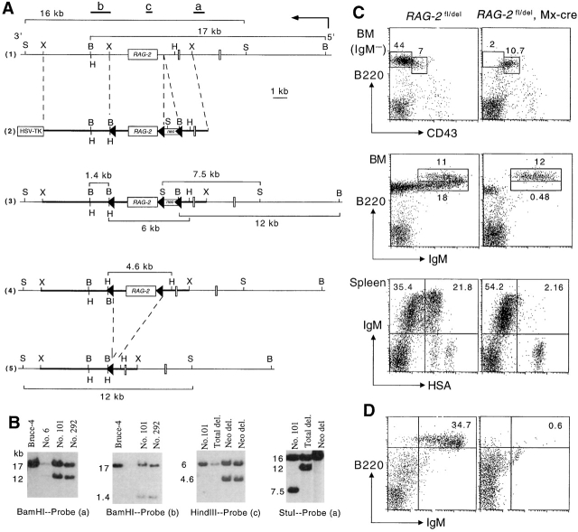
Figure 1.
Generation of RAG-2 fl/+ mice (A) targeting scheme. 1 Restriction map of the RAG-2 locus. Probes used to verify targeting events are indicated as a, b, and c together with the expected sizes of the restriction fragments. The restriction sites of XbaI (X), BamHI (B), HindIII (H), and StuI (S) are indicated. 2 Targeting vector construction. The loxP flanked neomycin resistance gene was inserted into a unique SalI site. A third loxP site was introduced downstream of exon 3. In this way, the entire coding sequence for RAG-2 protein was flanked by loxP sites (triangles). The structure of the targeted RAG-2 locus 3, the targeted locus after removal of the neomycin resistance gene 4, and the locus following deletion of the loxP-flanked exon 3 5 are shown, including sizes of diagnostic restriction fragments. (B) Southern blot analysis of ES cell clones. Genomic DNA from wild-type Bruce-4 cells and clones 6, 101, and 292 were digested with BamHI and hybridized with probe (a) to verify the targeting event or with probe (b) to detect cointegration of the third loxP site. Probe (c) together with HindIII digestion and probe (a) together with StuI digestion were used to distinguish subclones that had deleted only the neomycin resistance gene or the neomycin resistance gene and the entire loxP flanked fragment after transient transfection of the recombinants with a Cre-expressing plasmid. Numbers on the left side of the blots indicate fragment sizes in kb. (C) Block of B cell development upon the induction of RAG-2 deletion. Adult mice carrying or not carrying the Mx-cre transgene were injected with Poly(I)·Poly(C) and BM and spleen cells analyzed by FACS® 2 wk later. Numbers indicate the percentage of total lymphocytes. (D) BM cells of Poly(I)·Poly(C) treated RAG-2 fl/del, Mx-cre mice have no B cell reconstitution potential as shown by transfer into sublethally irradiated RAG-1 −/− mice. A flow cytometric analysis of spleen cells isolated from recipients that had received control (left) or RAG-2–deficient (right) BM cells 4 wk earlier is depicted.

