
Figure 3.
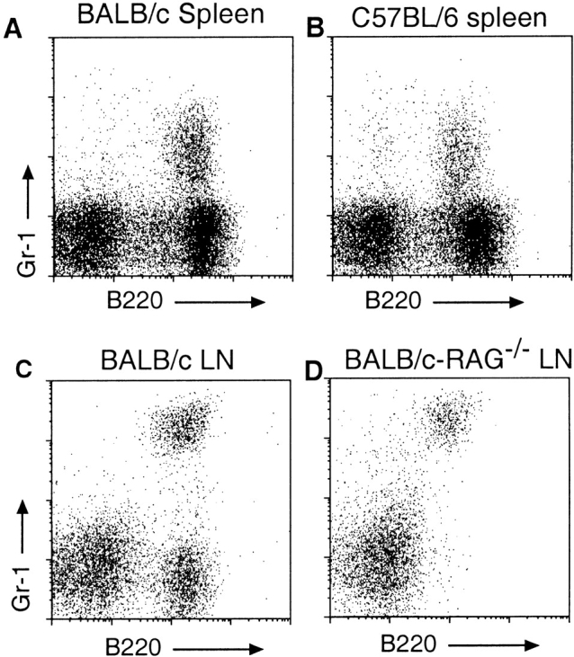
Presence of low density B220+Gr-1+ cells in BALB/c, C57BL/6, and Rag− /− mice. Low density cells were prepared from the indicated organs of mice as described in the legend to Fig. 1, stained with the indicated mAbs, and gated on low autofluorescent, 7-AAD− cells. (A and B) Presence of B220+Gr-1+ cells in the spleens of BALB/c and C57BL/6 mice, respectively. (C and D) Frequency of B220+Gr-1+ cells in the LNs of BALB/c and BALB/c-Rag−/− mice. The intensity of Gr-1 staining in top and bottom panels are not directly comparable due to differences methodology.