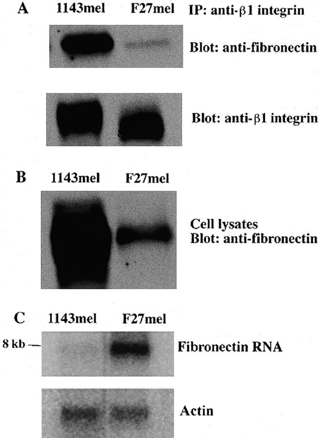
Figure 6.
Western and Northern blot analyses of FN in different tumor cell lines. (A) Tumor cell lysates of 1143mel and F27mel were first immunoprecipitated with an anti-β1 integrin. The immunoprecipitated proteins were separated on an SDS-PAGE. After the transfer to membrane, proteins were detected with anti-FN or anti-β integrin Abs. (B) Whole tumor cell lysates of 1143mel and F27mel cells were separated by SDS-PAGE and analyzed with the anti-FN Ab to determine the total FN protein in the tumor cell lysates. (C) Northern blot analysis of total RNA isolated from 1143mel and F27mel cells. Hybridization of blots with the probe of FN detected an 8-kb band in 1143mel and F27mel tumor cells. An actin probe was used to verify that equal amounts of total RNA were loaded for each well. FN-specific RNA in F27mel cells was at least three- to fourfold higher than that in 1143mel cells.

