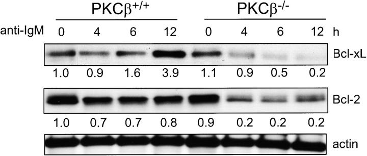
Figure 2.
Reduced expression of Bcl-xL and Bcl-2 after IgM cross-linking on B cells of PKCβ−/− compared with control mice. Splenic B cells were isolated from PKCβ−/− and PKCβ+/+ mice and incubated with 10 μg/ml anti-IgM for the indicated time (h). Bcl-xL and Bcl-2 protein expression was analyzed by Western blot analysis. Equal protein loading was controlled by anti-actin Western blotting. For quantification, band intensities were first normalized to the respective actin signal and then calculated as fold-change relative to unstimulated PKCβ+/+, which was set to 1.0.

