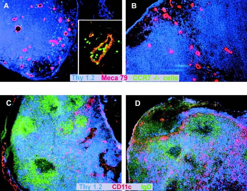
Figure 3.

(A and B) CCR7−/− lymphocytes are able to migrate through HEVs into secondary lymphoid tissue after FTY720 treatment. Splenocytes from a CCR7−/− donor animal were labeled with CFDA-SE. Cells were treated ex vivo with FTY720 (0.5 μM, 37°C, 2 h) or incubated in medium only (37°C, 2 h) and injected into wild-type recipient mice. After 2.5 h, LNs were obtained by dissection and cryostat sections were prepared. HEVs were stained with MECA-79 mAb and counterstaining was against CD90 (Thy1). (A) FTY720-treated CCR7−/− cells migrate through HEVs (insert, 40× original magnification) and into surrounding tissue (large panel, 10× original magnification). (B) Untreated CCR7−/− cells do not migrate into tissue surrounding HEVs (10× magnification). Representative results of independent experiments with four recipient animals each (treated and untreated) are shown. (C and D) Increased numbers of T cells migrate into PLNs of CCR7−/− mice but their tissue distribution is not altered after FTY720 treatment. CCR7−/− mice were treated with FTY720 (2 μg/ml) (C) or vehicle only (D) for 10 d. LNs were obtained by dissection and cryostat sections were prepared. T cells were stained with anti-Thy 1-Cy5, B cells were stained with anti-IgD-FITC, and DCs were stained with CD11c-biotin/streptavidin-Cy3.
