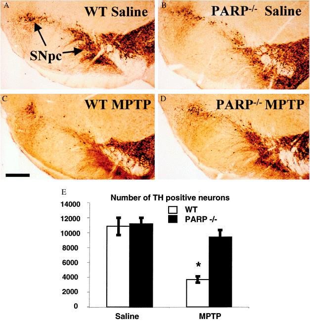
Figure 2.
DA neurons from PARP−/− mice are resistant to MPTP neurotoxicity. TH immunostaining of representative midbrain sections 7 days after MPTP adminstration from (A) saline-injected WT, (B) saline-injected PARP−/−, (C) MPTP-injected WT, and (D) MPTP-injected PARP−/− mice. (E) A significant reduction of TH-immunopositive neurons is seen in the WT mice receiving MPTP (n = 5) compared with saline controls (n = 8) (∗, ANOVA with Fisher post hoc, P < 0.0001 WT MPTP vs. saline). No statistical difference is seen between saline controls and PARP−/− (n = 4) 1 week after MPTP administration (ANOVA). Counts of Nissl-stained neurons in midbrain yielded similar results (data not shown).

