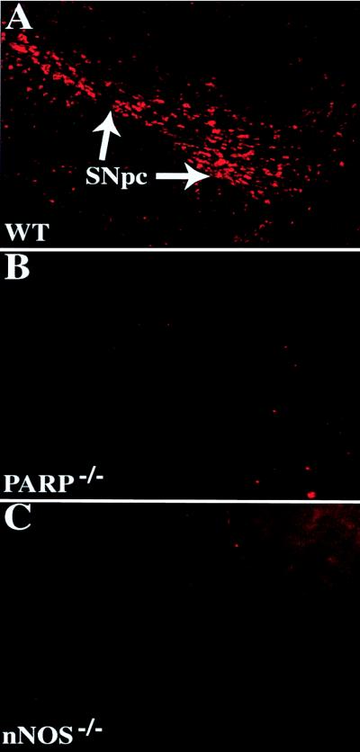
Figure 4.
PARP is activated in DA neurons after MPTP intoxication. Immunohistochemical staining with an anti-poly(ADP-ribose) antibody (pseudocolored in red) in the ventral midbrain. (A) WT after MPTP delivery demonstrates intense and specific staining of DA neurons. (B) PARP−/− midbrains are devoid of immunostaining. (C) nNOS−/− mice lack poly(ADP-ribose) formation after MPTP. Poly(ADP-ribose) is not detectable in saline-injected animals (data not shown). These images were obtained from animals 4 hr after the fourth injection of MPTP. These experiments have been replicated three times with similar results.

