Figure 6.
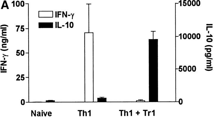
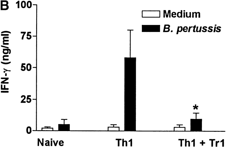
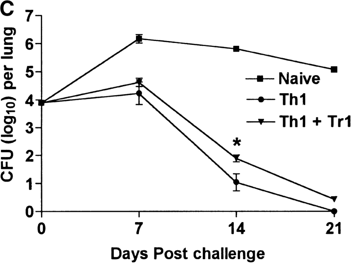
Tr1 clones suppress B. pertussis–specific IFN-γ production in vitro and in vivo. (A) Spleen cells from B. pertussis convalescent mice 6 wk after challenge (designated Th1) were cultured at 2 × 10⁶/ml with antigen (B. pertussis sonic extract, 5 μg/ml) alone or with Tr1 clone TEK.1 (105/ml) and PRN (5 μg/ml). Spleen cells from naive mice served as a control. Supernatants were removed after 3 d and tested for IFN-γ and IL-10 by immunoassay. (B and C) Spleen cells from naive or convalescent mice (designated Th1) were transferred into sublethally irradiated BALB/c mice (2 × 10⁷ per mouse) alone or with Tr1 clone TEK.1 (2 × 10⁵ per mouse). Mice were challenged with B. pertussis and killed 7, 14, and 21 d later. (B) Spleen cells were isolated after 7 d and stimulated with B. pertussis antigen and IFN-γ concentrations determined in supernatants 3 d later. (C) Lungs were removed and the number of viable B. pertussis determined by performing CFU counts. Results are mean (SD) for four mice per group at each time point. *P < 0.05, Th1 vs. Th1 plus Tr1. The results shown in A are representative of two experiments and due to the large numbers of cells required the experiment shown in B and C were performed once.


