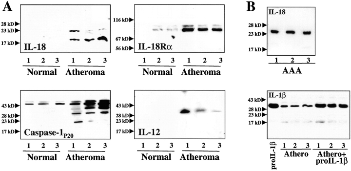
Figure 3.
Human atherosclerotic lesions contain immunoreactive IL-18 and IL-18Rα. (A) Extracts (50 μg/lane) of nonatherosclerotic arterial specimens (Normal) as well as atherosclerotic lesions (Atheroma) were applied to SDS-PAGE and subsequent Western blot analysis using either an anti–IL-18, anti–IL-18Rα, anti–caspase-1P20, or anti–IL-12P40 antibody. (B) As control for protein degradation, (top) protein extracts of abdominal aortic aneurysm (AAA) were applied to Western blot analysis for IL-18 or (bottom) exogenous recombinant IL-1β precursor (300 ng/mg tissue) was added to extracts of atherosclerotic tissue. The positions of the molecular weight markers are indicated on the left. Analysis of tissue obtained from a total of five nonatherosclerotic as well as seven atherosclerotic specimens showed similar results.

