
Figure 5.
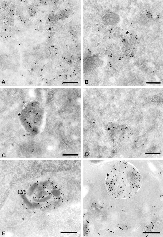
Characterization of the multivesicular compartments induced after BCR stimulation. Ultrathin cryosections of IIA1.6 cells stimulated for 30 min were double immunogold-labeled with the rat monoclonal M5.114 antibody and a polyclonal anti-invariant chain antibody (A), a polyclonal anti-FITC antibody (B) or a rat monoclonal anti-Lamp1 antibody (D). The same cells were also single immunogold-labeled with a rabbit polyclonal anti-Igα antibody (C). In D, a biotinylated M5.114 antibody was used to avoid cross-reactions. In E and F, ultrathin cryosections of unstimulated IIA1.6 cells (E) or cells stimulated for 30 min (F) were double immunogold-labeled with the M5.114 antibody (PAG 10) and a biotinylated anti–H2-M antibody detected with an anti-biotin antibody (PAG 15). (A) Note the presence of the invariant chain (PAG 15) in the class II-rich multivesicular compartments (stars). (B) The FITC-coupled Igs (PAG 15) clearly accumulate in the class II molecule-containing multivesicular compartments (PAG 10). (C) The multivesicular MIICs also contain Ig α (PAG 10) (star). (D) Both Lamp1 (PAG 15) and MHC class II (PAG 10) are present in the multivesicular MIICs. (E) In unstimulated cells, most of the H2-M was present in compartments containing internal electron-dense membranes (Lys). (F) In cells stimulated for 30 min, H2-M was detected in characteristic class II molecule-containing multivesicular compartments (star). Bars, 200 nm.