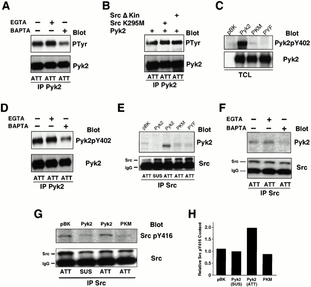
Figure 5.
Pyk2 is autophosphorylated at Y402 after adhesion and associates with Src via the phosphorylated Y402. (A) 293-VnR cells transiently transfected with 5 μg of Pyk2 were treated with calcium chelators BAPTA and/or EGTA, and then replated on serum-coated plastic and allowed to attach to vitronectin-coated plastic for 30 min. Pyk2 IPs from lysates were Western blotted for P-Tyr (top), and then reprobed with anti–Pyk2 (bottom). (B) 293-VnR cells were transiently transfected with Pyk2 (5 μg) or cotransfected with Pyk2 (5 μg) and Src kinase-inactive mutants (10 μg). Pyk2 IPs from lysates were probed with P-Tyr antibodies (top). Membrane was stripped and reprobed with Pyk2 (bottom). (C) Pyk2 and Pyk2 mutants Y402F (PYF) and K457A (PKM) were transiently expressed in 293-VnR cells and total cell lysates (TCL) were blotted with an antibody specific for Pyk2 phosphorylated residue 402 (top). (Bottom) Transfected proteins. (D) Cells were processed as in A and Pyk2 IPs were blotted with an antibody specific for Pyk2 phosphorylated residue 402 (top). (Bottom) Pyk2 blot demonstrating equal loading. (E) 293-VnR cells were transiently transfected with wild-type Pyk2, kinase-dead Pyk2 (PKM) or Pyk2Y402F (PKF). The transfected cells were harvested, and then kept in suspension (SUS) or replated on serum-coated plastic (ATT) as in Fig. 4 E. Association of Src with mutant Pyk2 proteins was analyzed by Western blotting Src immunoprecipitates with Pyk2 antibodies (top). (Bottom) The membrane reprobed with Src antibodies. (F) Src IPs from cells treated with either EGTA or BAPTA were blotted with Pyk2 antibodies (top). (Bottom) The membrane reprobed with anti–Src. (G) 293-VnR cells transfected with 5 μg of Pyk2 or dominant-negative kinase-dead Pyk2 (PKM) were either kept in suspension or replated on vitronectin coated dishes for 30 min. Lysates were immunoprecipitated with anti–Src antibodies and probed with Src pTyr-416 antibody (top), which detects the activated form of Src. The membrane was stripped and reprobed with anti–Src (bottom). (H) Src kinase activity was quantified as an increase in pY416 Src using Scion Image1.62 C program.

