
Figure 2.
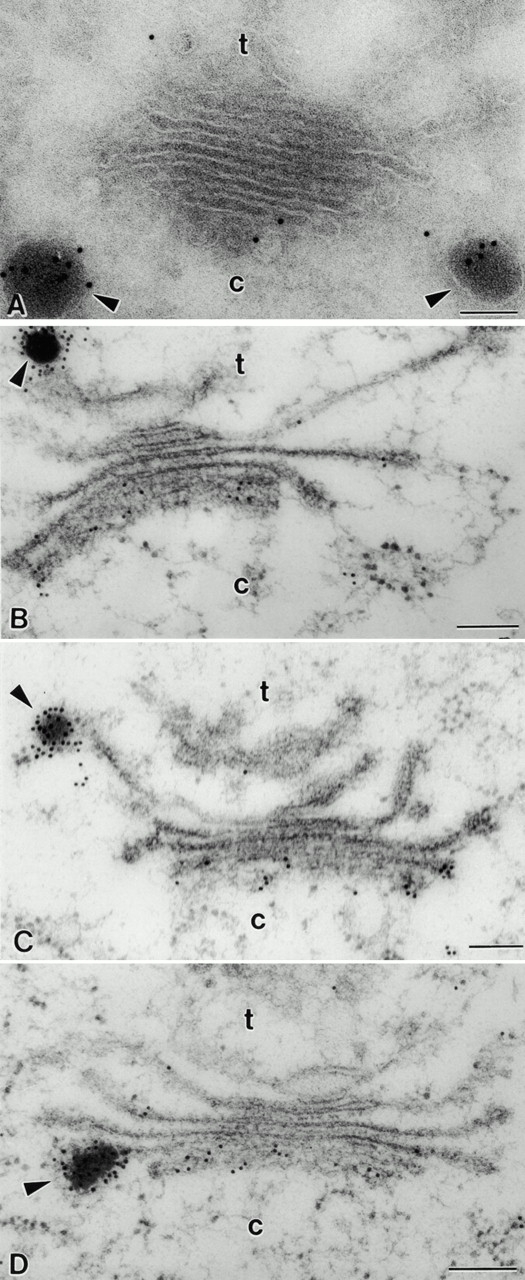
Immunogold staining of pea cotyledon Golgi stacks with antibodies against vicilin. (A) Cryosectioned sample. Background labeling is very low. DVs are heavily labeled, and the label in the Golgi stack is restricted to the cis-cisternae. (B, C, and D) HPF sample. Labeling of the cisternae reveals a steep gradient from the cis to the trans side of the Golgi. DVs are seen attached to the TGN (B), the trans-cisternae (C), and median cisternae (D). c, cis; t, trans. Bars, 100 nm.