Figure 4.

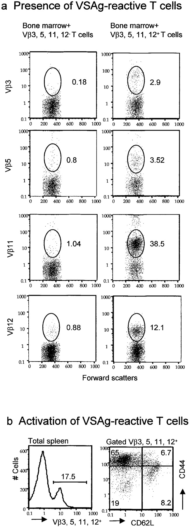
Presence and pathogenicity of VSAg-specific T cells rescued by perinatal treatment with anti-B7 mAbs. Lethally irradiated BALB/c mice (900 rad) were adoptively transferred with 107 per mouse of syngeneic bone marrow cells and 3 × 10⁶ per mouse of thymocytes, enriched for their expressing either Vβ3, Vβ5, Vβ11, or Vβ12. As control, 3 × 10⁷ thymocytes depleted of the Vβ3-, Vβ5-, Vβ11-, and Vβ12-expressing cells were also adoptively transferred together with syngeneic bone marrow cells. 3 wk after adoptive transfer, the recipient mice were killed and the spleen cells were analyzed. (a) Large numbers of VSAg-specific T cells were found in syngeneic VSAg-expressing mice that received bone marrow plus Vβ3, Vβ5, Vβ11, or Vβ12+ cells (right), but not those that received bone marrow plus Vβ3, Vβ5, Vβ11, and Vβ12− cells (left). Data shown were gated CD4+ cells from spleen cells and are representative of the CD4 T cells from all five mice analyzed. The numbers in the panels are the percentage of cells expressing given Vβ among CD4+ T cells. (b) The majority of the VSAg-reactive T cells expressed markers of antigen-experienced T cells. Spleen cells in the chimera mice were stained with FITC-conjugated mAbs specific against Vβ3, Vβ5, Vβ11, or Vβ12. Cychrome–anti-CD44 and PE–anti-CD62L mAbs were added. Data shown in the left panels are histograms for distribution of Vβ3-, Vβ5-, Vβ11-, or Vβ12-expressing cells. The data shown in the right are dot plots depicting the expression of CD44 and CD62L.
