
Figure 7.
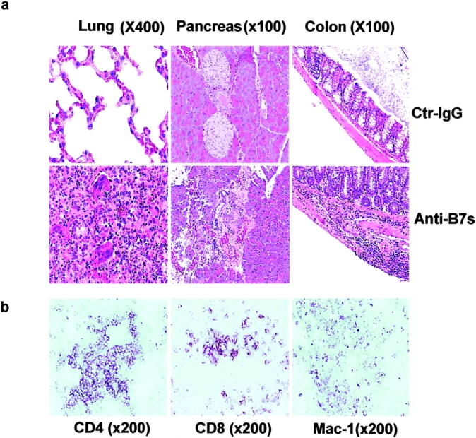


Perinatal blockades of B7-1– and B7-2–induced T cells that cause chronic autoimmune inflammation in adult RAG-1−/− recipients. Spleen T cells were isolated from 3-wk-old female C57BL/6j mice that had received either anti-B7 mAbs or control IgG and injected intraperitoneally (2 × 10⁷ per mouse) into 6-wk-old, syngeneic female RAG-1−/− mice (n = 5 per group). The recipient mice were killed 3 mo after adoptive transfer. (a) H&E staining of lung, liver, and colon sections. In the anti–B7-treated group, multinucleated giant cell granulomas, characteristic of chronic inflammation, were present in the lung. Pancreatic acini were infiltrated by mononuclear cells, and neolymphoid follicles were observed in some mice. The villi of the small intestine were densely infiltrated by lymphocytes (unpublished data), and in some areas inflammatory cells were present in the submucosa and muscularis externa of the colon. The sections presented are from a representative mouse from each group. All mice that received T cells from anti–B7-treated mice developed severe inflammation in the lung, liver, pancreas, and intestine, whereas the recipients of control IgG-treated T cells were histologically normal, with occasional foci or inflammation in the lung. (b) Immunohistochemical analysis of lung sections using anti-CD4, anti-CD8 and anti–Mac-1 antibodies. The pictures were taken from the same anatomic location of three consecutive sections. (c) Numbers and phenotypes of T cells in the spleens and lymph nodes of RAG-1−/− mice that were adoptively transferred with splenic T cells from anti–B7-treated mice. Splenocytes and lymph node cells were isolated, counted, stained with FITC-conjugated anti-TCRαβ, PE-conjugated anti-CD4, and Cychrome–conjugated anti-CD8 (left and middle), or FITC-conjugated anti-TCRαβ, PE-conjugated anti-CD62L, and Cychrome–conjugated anti-CD8, and were analyzed with flow cytometry. The number of CD4−CD8− T cells were obtained by subtracting the CD4+CD8− and CD8+CD4− T cells from the TCR+ T cells. (d) Intracellular cytokine expression in the T cells. Data shown are expressions of IFN-γ from gated CD4, CD8, and CD4−CD8− lymphocytes. No alteration in the number of IL-2–, IL-4–, and IL-10–producing cells was observed (unpublished data). Data shown were representative of two to three independent experiments.