
Figure 3.
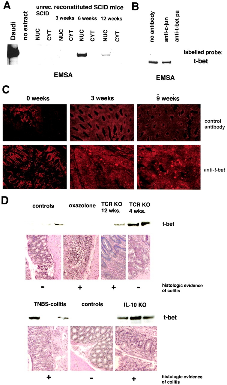
Increased expression of T-bet in Th1- but not Th2-mediated animal models of chronic intestinal inflammation A. Analysis of T-bet expression in C.B.-17 SCID mice reconstituted with 106 CD62L+ CD4+ T cells from Balb/c mice. EMSA for T-bet using nuclear (NUC) and cytoplasmic (CYT) extracts from spleen cells of unreconstituted and CD62L+ CD4+ T cell reconstituted SCID mice at indicated time points. SCID mice showed clinical signs of colitis 8–9 wk after T cell transfer. (B) Supershift assay for T-bet using nuclear extract from spleen cells of SCID mice 6 wk after reconstitution with CD62L+ CD4+ T cells from Balb/c mice. (C) Immunohistochemical analysis of the colon of SCID mice for T-bet expression at indicated time points after reconstitution with CD62L+ CD4+ T cells. T-bet expressing cells in the LP were noted as soon as 3 wk after T cell transfer. (D) Expression of T-bet in extracts from LPMCs in Th1- and Th2-mediated animal models of chronic intestinal inflammation. Whereas increased T-bet expression by Western blot analysis was noted in Th1-mediated models such as TNBS-induced colitis in Balb/c mice and colitis in 4–12-wk-old IL-10 knockout mice, both oxazolone-induced colitis and TCR-α−/− μ−/− colitis were associated with normal or reduced T-bet expression in LPMCs.