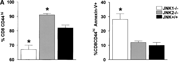
Figure 4.
During the acute infection, the number of splenic CD8+ T cells CD44hi is lower in JNK1−/− mice than in JNK2−/− and JNK+/+ mice, and a greater proportion of these cells from JNK1−/− undergo apoptosis. JNK1−/−, JNK2−/−, and JNK+/+ animals were injected intraperitoneally with LCMV ARM 2 × 10⁵ PFU. 8 d after infection, spleen were processed to make single cell suspension devoted of red blood cells and stained directly for surface markers: CD4, CD8, and CD44 then annexin-V and 7AA-D were added. Data represent average values obtained from four animals per group and are representative of two independent experiments. The percentage of CD8+ or CD4+ that were CD44hi are shown in the left, that were annexin V+ cells among the CD8CD44hi or CD4CD44hi on the right. (A) CD8+ T cells. (B) CD4+ T cells. *P < 0.05 compared with JNK+/+.


