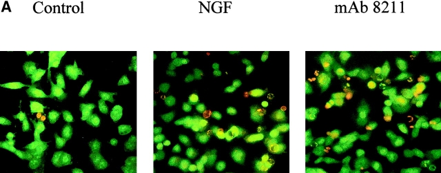
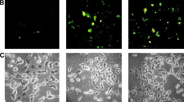
Figure 6.
Epifluorescence microscopic analysis of cell damage by NGF and mAb 8211. BEp75 cells untreated and treated for 24 h with NGF (10 nM) or mAb 8211 (5.0 μg/ml) (A) stained with OA plus EB (for details see Fig. 2), or (B) with Annexin V-FITC plus propidium iodide. (C) Phase contrast images corresponding to those in B.


