Figure 1.
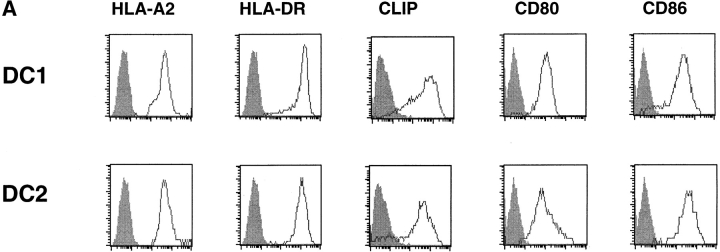
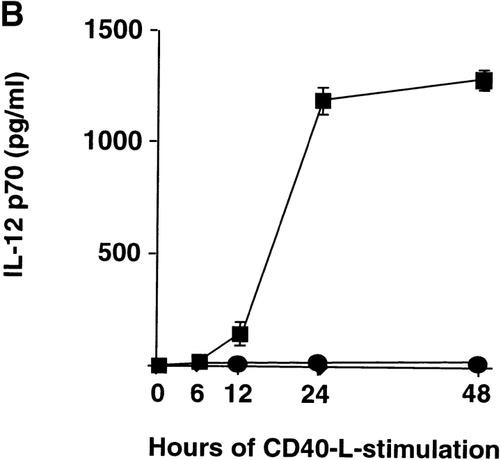
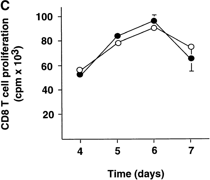
DC1 and DC2 express similar levels of MHC products and costimulatory molecules, but differ in their ability to produce IL-12. (A) Cell surface expression of HLA-A2, HLA-DR, class II–associated peptides (CLIP), CD80, and CD86 on DC1 and DC2. (B) IL-12p70 in the supernatants of DC1 generated in a 5-d culture with GM-CSF and IL-4 (▪) and DC2 generated in a 5-d culture with IL-3 (•) stimulated at 100,000 cells/ml with CD40 ligand–transfected L cells for 6, 12, 24, and 48 h. Mean value ± SD of two independent experiments are represented. (C) DC1 and DC2 induce strong proliferation of naive CD8 T cells. Naive CD8 T cells were stimulated with allogeneic CD40 ligand–activated DC1 (•) and DC2 (○) and proliferative responses were assessed after 4, 5, 6, or 7 d. Results are expressed as mean cpm ± SD of triplicate wells. Background [3H]thymidine incorporation of CD8 T cells cultured in the absence of DC remained <1,000 cpm at all time points.


