
Figure 8.
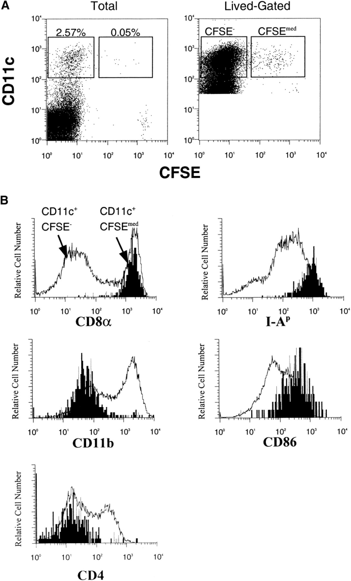

Splenic DCs containing transferred proteins express high levels of CD8α, I-A, and CD86. Purified HEL-specific transgenic B cells on the C57Bl/6 (H-2bb) background were labeled with CFSE, loaded with HEL ex vivo, and adoptively transferred into unmanipulated B10.BR (H-2kk) recipients. (A) Data were first collected for total live cells, and then subsequent data were collected using lived-gating on CD11c+ to collect sufficient events for analysis. (B) Lived-gated files were gated in the CD11c+, CFSE− and CD11c+, CFSEmed populations identified in the Figure and were analyzed for the expression of surface markers by flow cytometry. Data were gated as in Fig. 6. The data are representative of three independent experiments. (C) Stable surface phenotype of CD11c+CFSEmed early after donor cells injections. B6 splenocytes (H-2bb) were labeled with the dye CFSE and adoptively transferred into B10.BR recipients (H-2kk) by intravenous injection. The recipient mice were killed 2–6 h after the injections. Spleens were prepared and analyzed by flow cytometry. The CD11c+ CFSE− and CD11c+ CFSEmed populations were identified, then analyzed for the expression of surface markers. Data were gated as in the Figure.