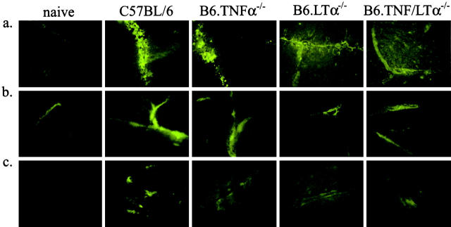
Figure 3.
Comparative immunohistochemical analysis of brain sections taken from P. berghei-infected mice when C57BL/6 and B6.TNFα−/− mice developed CM (day 7 after infection). Brain cryosections (6 μM) were stained with (a) anti-P. berghei antibodies, (b) anti-ICAM-1, and (c), anti CR3 (5C6) mAbs with fluorescent secondary reagents. Original magnification is ×400. The samples are representative of material from four experiments.

