
Figure 7.
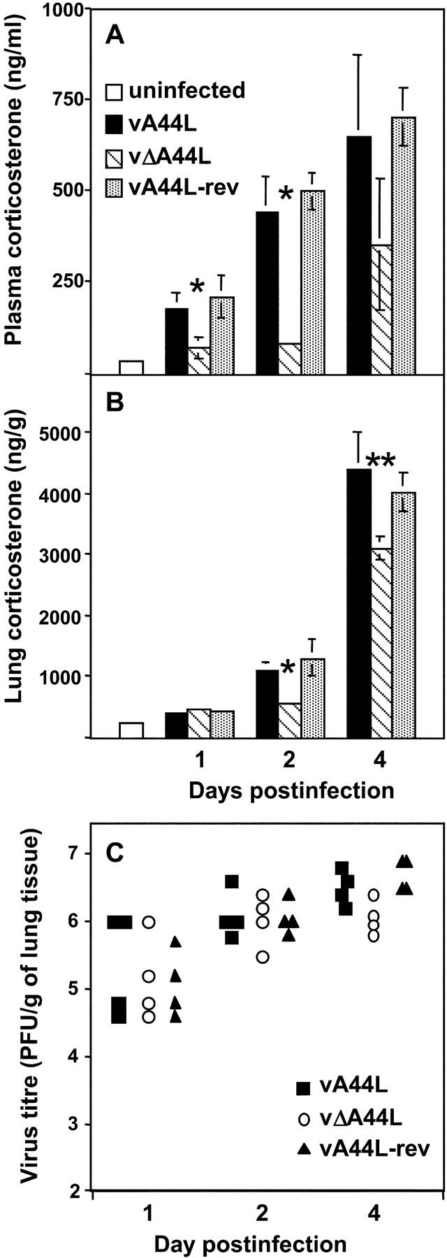
Corticosterone levels in plasma and lungs after intranasal infection with VV. Corticosterone levels were measured in (A) plasma and (B) lung extracts collected from BALB/c mice under low stress conditions (samples were obtained within 4 min of handling) at the indicated times after intranasal infection with 105 PFU of vA44L, vΔA44L, or A44L-rev. Lung extracts were prepared as described in Materials and Methods. Data represent mean ± SEM of four or five mice per time point and are expressed as ng/ml of plasma or as ng/g of lung tissue. Columns marked with an asterisk represent corticosterone levels from vΔA44L-infected mice that were significantly different to those from vA44L- and vA44L-rev-infected mice. *, P < 0.05, **, P < 0.02. (C) Titers of infectious virus in the lungs of mice after infection with 105 PFU of VV. Virus titers were determined by plaque assay on BS-C-1 cells and are expressed as PFU/g of lung tissue.