
Figure 2.

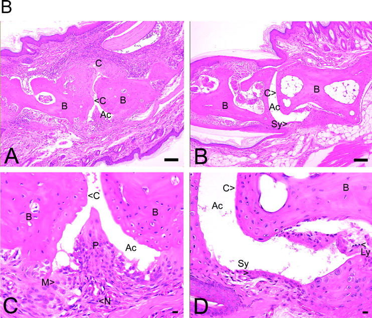
(A) Summary of histopathological grades for synovial hyperplasia, inflammatory cells, pannus formation, ankylosis, and fibrillation of all joints of cPLA2α-deficient and wild-type mice. Significant differences to the wild-type group are marked with *** for P < 0.0001, ** for P < 0.001, and * for P < 0.01 (two-tailed t test). Concerning ankylosis, t test was not applicable because all cPLA2α-deficient mice scored zero. (B) Histopathology of hematoxylin and eosin–stained joints of front paws of cPLA2α-deficient and wild-type mice. In A, the joints of wild-type mice most frequently showed severe pathology with cartilage erosion, synovial inflammation, and formation of invasive pannus (P). (B) The pannus (see close-up in C) was comprised of a mixture of monocyte/macrophages (M) and neutrophils (N). In B, the majority of examined joints of cPLA2α-deficient mice were normal in appearance, with smooth intact articular cartilage. In D, the close up of B showed few infiltrating cells, e.g., lymphocytes (Ly). Ac, articular cavity; B, bone. original magnification: ×10 for A and B, bar = 0.1 mm; ×40 for C and D, bar = 0.01 mm.