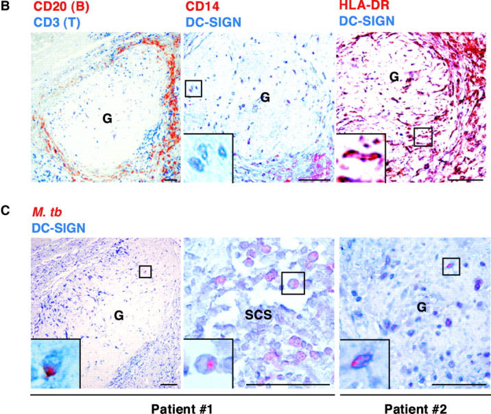
Figure 3.

DC-SIGN expression on lung DCs (LDCs) and in lymph nodes (LNs). (A) LDCs are HLA-DR+ and express DC-SIGN, CR3, and MR. Surface expression of HLA-DR, DC-SIGN, CR3/CD11b, and MR on LDCs from a noninfected patient was assessed by flow cytometry using the appropriate cytochrome-conjugated mAbs. (B) DC-SIGN expression in the LN from a patient with tuberculosis (G, granuloma). Left panel, CD3 (blue) and CD20 (red); middle panel, DC-SIGN (blue) and CD14 (red); right panel, DC-SIGN (blue) and HLA-DR (red). (C) Localization of M. tuberculosis–derived antigens in DC-SIGN+ cells in LNs from two patients with tuberculosis. DC-SIGN (blue) and M. tuberculosis (red) were immunodetected both in granulomas (G; left and right panels) and in nongranulomatous regions, including subcapsular sinuses (SCS; middle panel). In B and C, bars represent 0.5 mm and squares represent areas shown at higher magnification at the single cell level in the insets. Staining of the samples with IgG2a (1B10 isotype control) or with a naive rabbit serum led to no detectable signal (data not depicted).

