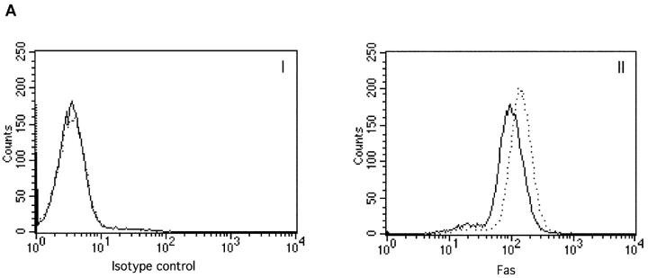
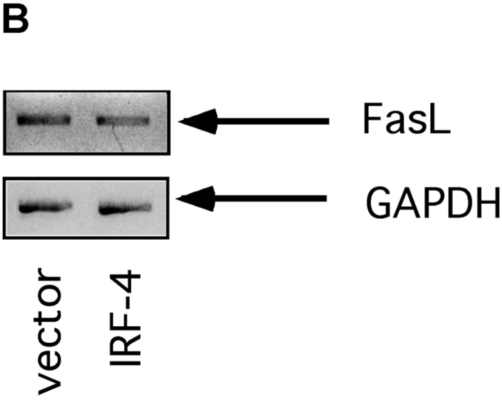
Figure 2.
Expression of Fas and FasL in control and IRF-4 transfectants. (A) Surface expression of the Fas receptor in Jurkat transfectants. Cells from control and IRF-4–transfected cells were stained with either a PE-labeled isotype-matched control Ab (I) or a PE-labeled anti-Fas IgG1 antibody (II). Cells were subsequently analyzed by flow cytometry. Control vector transfectants (solid lines); IRF-4 transfectants (dotted lines). (B) RT-PCR analysis for FasL expression in IRF-4 Jurkat transfectants. Total RNA was isolated from control and IRF-4–transfected cells, and RT-PCR was performed using primers specific for FasL (top) or GAPDH (bottom). PCR products were separated by electrophoresis on a 2% agarose gel.


