Figure 8.
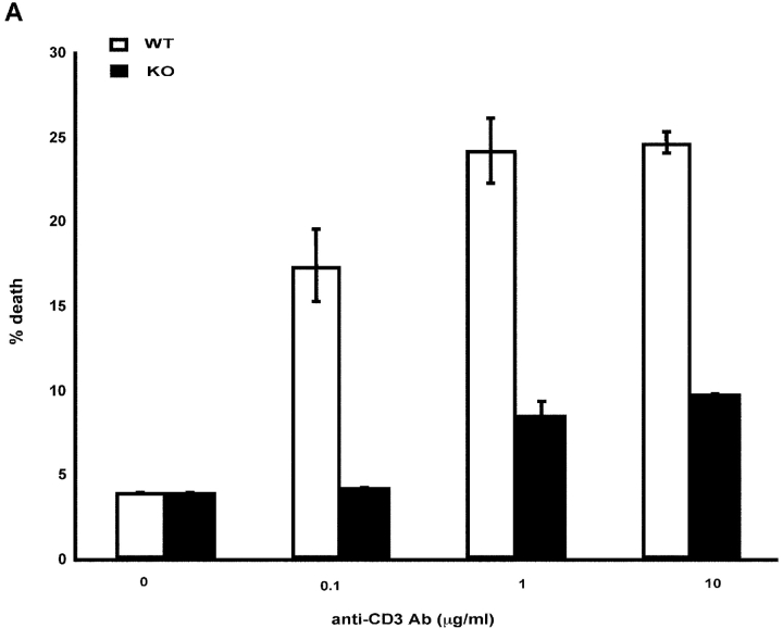
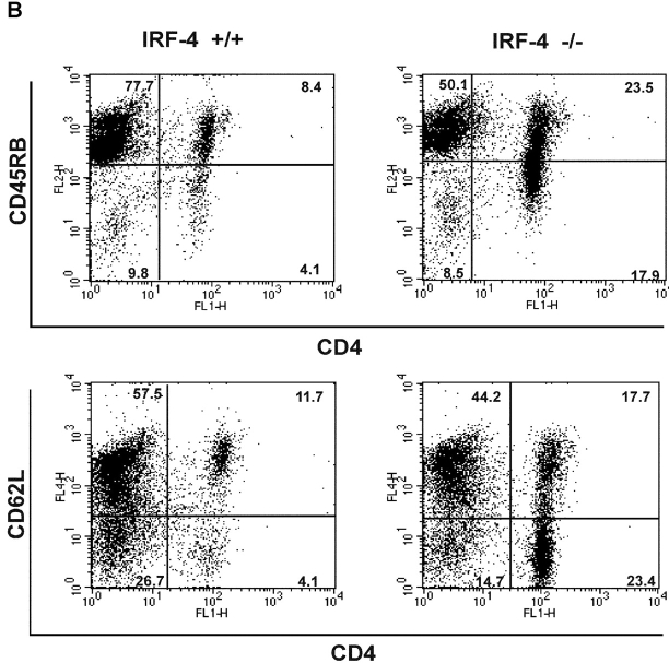
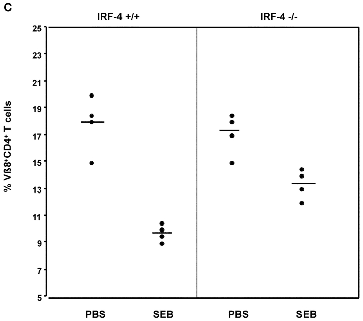
IRF-4–deficient mice exhibit impaired AICD and an accumulation of CD4+ T cells with an “experienced” phenotype upon aging. (A) AICD. Purified naive CD4+ T cells were activated with 1 μg/ml anti-CD3 plus IL-2 for 3 d, and then harvested. The cells were restimulated with either IL-2 alone or IL-2 + anti-CD3 at the indicated doses for 24 h. The percentage of apoptotic cells was determined by quantification of the sub-G0 population by FACS®. Each assay was conducted in duplicate. The experiment is representative of three separate experiments. (B) Flow cytometric analysis of spleen cells from 12–15-wk-old wild-type and IRF-4–deficient mice. Single cell suspensions of splenocytes were stained with anti-CD4 versus anti-CD45RB (top) or anti-CD4 versus CD62L (bottom). Percentages of positive cells within each quadrant are indicated. (C) Mice (four per treatment) were injected with either PBS or SEB. After 10 d, the percentage of Vβ8+CD4+ (SEB responsive) T cells in lymph nodes was determined by flow cytometry. Each symbol represents an individual mouse. Bars represent the mean of each group of four mice. The number of Vβ6+CD4+ (SEB unresponsive) T cells was also determined by flow cytometry as a control, and it was found not to be affected by SEB treatment in either wild-type or IRF-4–deficient mice (unpublished data).


