
Figure 8.
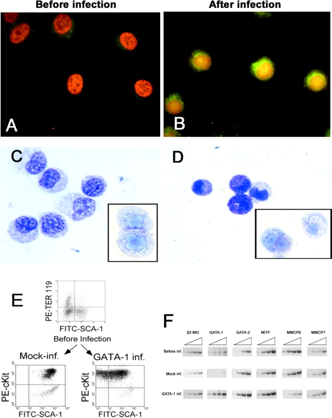
Infection of GATA-1low BMMC with the PGK-GATA-1 retrovirus increases the level of GATA-1 expression and restores in vitro maturation potential. (A and B) GATA-1–specific immunofluorescent staining of BMMC either before or 17 d after their coculture with the PGK-GATA-1–producing cell line, as indicated. Cell nuclei are counterstained with DAPI. (C and D) Alcian blue staining of the same cells shown above. Toluidine blue staining of representative cells are presented in the inserts. ×100 in all of the cases. (E) Immunophenotype of day 7 BMMC before infection and 17 d after the 48-h coculture with either NIH 3T3 (Mock-inf.) or the PGK-GATA-1–producing cell line (GATA-1 inf.). Day 7 BMMC were immunophenotyped with TER-119/SCA-1 whereas cocultured BMMC were immunophenotyped with c-Kit/Sca-1. (F) Semiquantitative RT-PCR analysis for the expression of β2 microglobulin (β2-MG, positive control), GATA-1, GATA-2, MITF, MMCP6, and MMCP7 genes in cells obtained after 7 d under BMMC-specific culture conditions (before infection) or 17 d after the BMMC had been cocultured with either NIH 3T3 (Mock inf.) or the PGK-GATA-1–producing cell line (GATA-1 inf.; the same cells as presented above). Each product was amplified for an increasing number of cycles (20, 25, 30, and 35), as indicated by the triangle on the top of the panel. Similar results were obtained in two additional experiments.