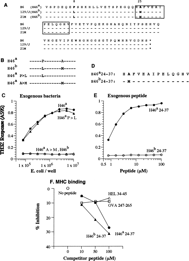
Figure 6.
Non-self at the H46 locus is defined by a single amino acid polymorphism. (A) The deduced amino acid sequences of H46 cDNA from the B6 (H46a), the congenic 21M (H46 b), and the parental 129/J (H46 b) mouse strains. The cDNA fragments corresponding to the B6-derived clone 224–26 were obtained by RT-PCR. The H46a antigenic peptide is boxed. (B) Constructs containing the H46a and H46b wild-type as well as single point mutations of each of the two polymorphic residues H46a P>L and H46a A>M subcloned into the bacterial expression vector pLEX. (C) TH3Z lacZ response to 129/J BMDCs fed with bacteria expressing the constructs shown in B. (D) Amino acid sequences of the synthetic peptide representing the H46a and H46b homologues. Bold letters indicate the single amino acid polymorphism between the B6 and 129 strains. (E) TH3Z lacZ response to the H46a and H46b peptides presented by 129/J-derived BMDCs. (F) The relative Ab MHC II binding property of H46a and H46b peptides compared with the Ak MHC II binding peptides HEL34–45 and OVA247–265. The data show the varying concentrations of indicated peptides cause competitive inhibition of the binding of the Eα peptide to the Ab MHC II on Ab-L cells. The Eα/Ab complex was detected with the Yae mAb by flow cytometry as described in Materials and Methods.

