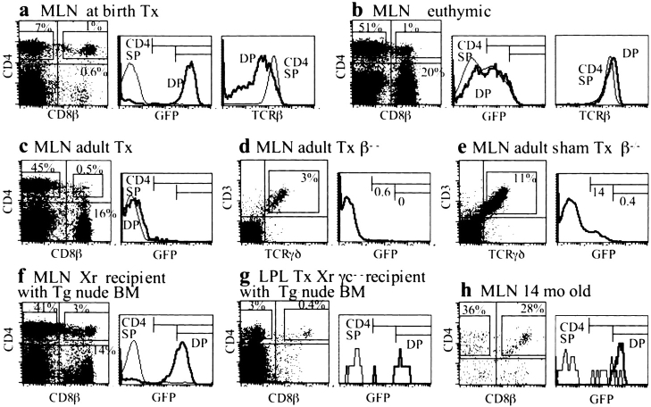
Figure 4.
Analysis of lymphopoiesis in various Tx or euthymic mice. (a) MLN cells, mouse Tx at birth (compare with Fig. 2 a). (b) MLN cells, 7-wk-old euthymic mouse, sham Tx (match of mouse with adult Tx shown in c). Presence of some DP with GFP and TCRβ expression similar to that of SP CD4+ cells (right panels). Note that many CD4+ cells are GFPL. (c) MLN, 7-wk-old mouse 2 wk after thymectomy. All DP and SP cells are GFP− (compare with b). (d and e) MLN cells from 8-wk-old Tg TCRβ−/− mice, Tx (d) or sham Tx (e) 2 wk earlier, used for easy study of γδ+ cells. GFPL cells are absent in the Tx mouse. (f) MLN cells of lethally irradiated non-Tg mouse reconstituted 18 d earlier with bone marrow cells from a nude Tg mouse. DP cells are GFPH. Note that MLN cell recovery was 10-fold less than that from normal mice. (g) Lymphocytes isolated from the LP of a Tx RAGγc−/− mutant mouse reconstituted 36 d earlier with nude Tg mouse bone marrow cells (note that some CD4+ GFP− cells, originating from the graft, are detectable). (h) MLN cells from a 14-mo-old mouse (cell recovery was ∼20-fold less than in younger mice). DP cells with GFP expression comparable to that of DP thymocytes.

