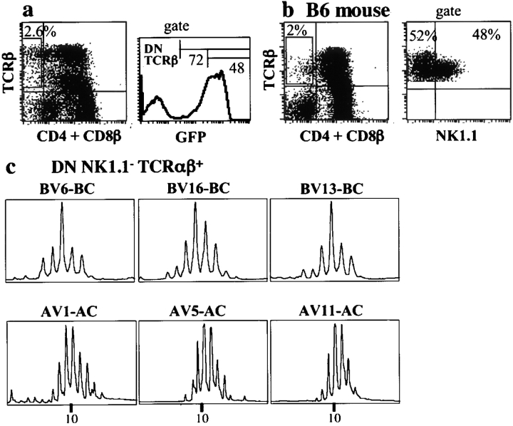
Figure 6.
Analyses of DN αβ1 thymocytes. Studies of (a) GFP content and (b) NK1-1 expression (in this last case, cells were from a B6 mouse because the Tg mice do not express NK1.1). (c) TCRα and β repertoires of DN NK1.1− thymocytes: polyclonal repertoires expressing chains different from that peculiar to NKT cells.

