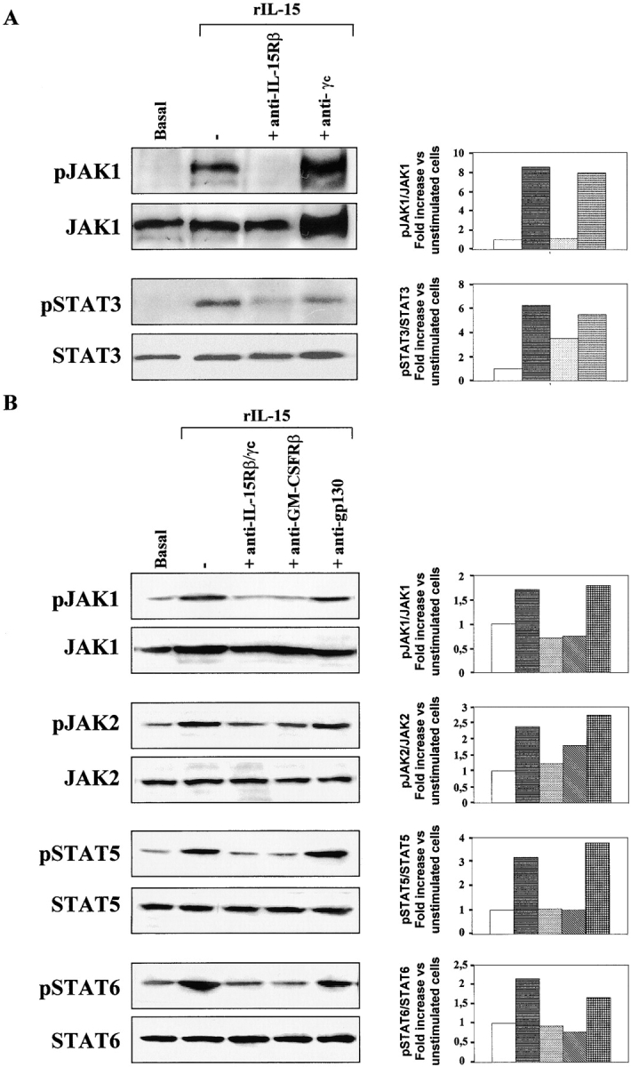
Figure 3.

IL-15 signal transduction: IL-15R/GM-CSFR cross talk in TF1β cells. Analysis of JAK/STAT signal transduction by Western blotting. TF1β cells were incubated with 10 ng/ml rIL-15 for 15 min at 37°C. Sister cultures were pretreated for 1 h with neutralizing anti–IL-15Rβ/γc, anti-GM-CSFRβ, or anti-IL-6R gp130 mAbs. Cell extracts were analyzed by Western blotting using anti-phospho-JAK1 (pJAK1), anti-phospho-JAK2 (pJAK2), anti-phospho-TYK2 (pTYK2), anti-phospho-STAT5 (pSTAT5), and anti-phospho-STAT6 (pSTAT6) antibodies. Membranes were then reprobed with antibodies recognizing the native proteins. To correct for possible variations in the amount of protein loaded, values are expressed as pJAK/JAK or pSTAT/STAT ratios. pJAK/JAK and pSTAT/STAT levels were determined by densitometry including correction for background (NIH Image software). Results are expressed as increases (e.g., two times) with respect to untreated cells. The data presented are representative of three independent experiments.
