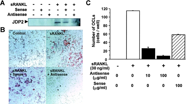
Figure 3.
Suppression of osteoclast formation by antisense oligonucleotide to JDP2 in RAW264.7 cells. (A) RAW264.7 cells (106 cells/well) cultured in DMEM containing 10% FCS in 60-mm-diameter dishes were treated with 10 µg/ml S-oligonucleotides (antisense, 5′-ATCATAGCAGGAGGAG-3′; sense, 5′-CTCCTCCTGCTATGAT-3′) in the presence of 100 ng/ml sRANKL for 24 h. Nuclear extracts were prepared and subjected to Western blot analysis using antibody for JDP2. (B) RAW264.7 cells (104 cells/well) cultured in DMEM containing 5% FCS in 96-well plates were treated with the sense or antisense S-oligonucleotide at 100 µg/ml in the presence of 30 ng/ml sRANKL for 4 d. The cells were fixed and stained for TRAP activity. (C) RAW264.7 cells were treated with indicated concentrations of the sense or antisense S-oligonucleotide in the presence of sRANKL (30 ng/ml) for 4 d, fixed, and stained for TRAP activity. TRAP-positive multinucleated cells containing more than three nuclei were counted as osteoclasts.

