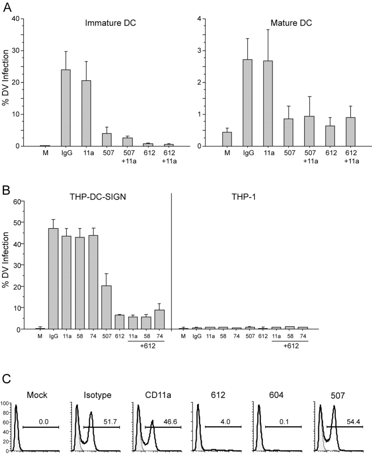
Figure 3.
Blocking studies of DV infection in DCs and THP-1. (A) Comparison of percent DV2 infection rate of immature (left) and mature DCs (right) in the presence and absence of anti–DC-SIGN mAbs (clones 120507-specific or 120612–cross-reactive), anti-CD11a, or an irrelevant matched isotype control. (B) Comparison of percent DV2 infection rate of THP DC-SIGN and THP-1 in the presence and absence of specific anti–DC-SIGN mAbs, anti-CD11a, anti-CD58, anti-CD74, or an irrelevant matched isotype control. Data are means (± SEM) of four independent experiments. (C) A representative blocking experiment (one of two) in the THP L-SIGN cells in the presence and absence of specific anti–DC-SIGN or L-SIGN mAbs (120604), anti-CD11a, or an irrelevant matched isotype control.

