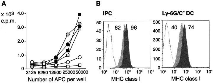
Figure 2.
Antigen presentation to HY-specific naive CD8 T cells by HY-transgenic IPCs and DCs. (A) Proliferation of naive CD8 T cells (5 × 10⁴) purified from HY-transgenic mice to IPCs (open symbols) or DCs (filled symbols) derived from uninfected or VSV infected H-2Db B6 males. VSV infection was performed as in Fig. 1. Proliferation was measured after 72 h. (B) Expression of H-2Db class I molecules in IPCs or DCs of uninfected mice (light gray histograms) or VSV infected mice (dark gray histograms). Open histograms show staining with a control antibody. Numbers indicate MFI.

