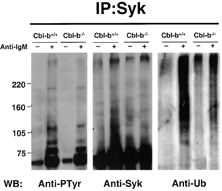
Figure 3.
The activation-induced ubiquitination of Syk is reduced in Cbl-b−/− B cells. Splenic B cells from Cbl-b−/− and Cbl-b+/+ mice were treated as in Fig 2, A–C, to cross-link the BCR and warmed to 37°C for increasing lengths of time. The cells were lysed, and Syk was immunoprecipitated and analyzed by SDS-PAGE and immunoblotting. Duplicate immunoblots were probed for either ubiquitin (anti-Ub) or phosphotyrosine (Anti-PTyr). The phosphotyrosine blot was stripped and reprobed for Syk (anti-Syk). The results for the 2-min time point are shown at which point ubiquitination of Syk was maximal. The result shown is representative of three independent experiments.

