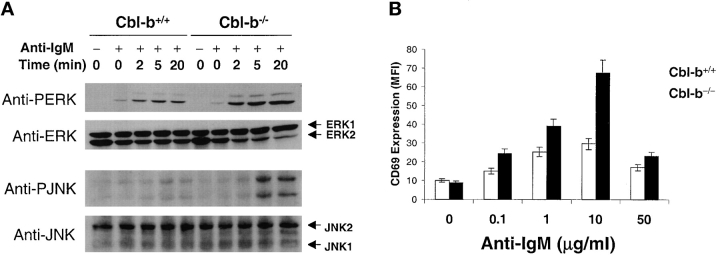
Figure 7.
ERK and JNK phosphorylation are prolonged and downstream CD69 expression increased in Cbl-b−/− B cells. (A) Purified splenic B cells were treated to cross-link the BCR as in Fig. 1. The cells were lysed and equal amounts of whole-cell lysates subjected to immunoblotting using Abs specific for phospho-ERK (anti-PERK) and phospho-JNK (anti-PJNK). The blots were stripped and reprobed for ERK1 and 2 and JNK1 and 2. (B) After BCR cross-linking the cells were incubated for 20 h at which time the surface expression of CD69 was quantified by flow cytometry. The results are the mean of duplicate cultures of one of three representative experiments.

