Figure 4.
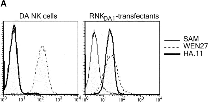
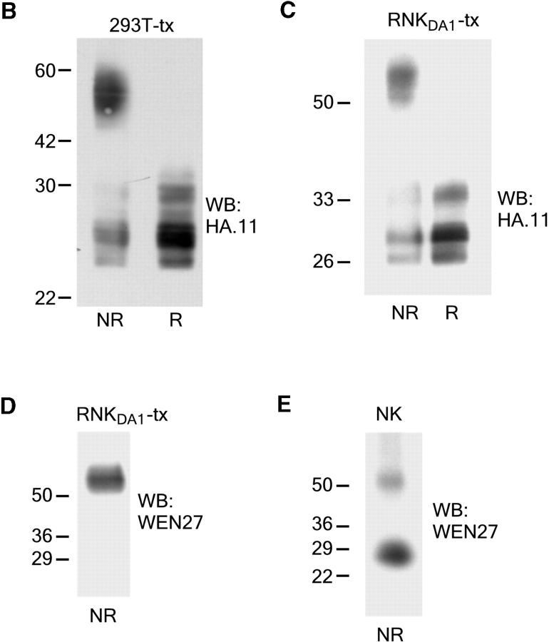
KLRE1 can be expressed as a dimer in the NK cell membrane. The IL-2–dependent RNKDA1 cell line was stably transfected with a KLRE1-HA expression construct. (A) RNKDA1–tx cells express KLRE1-HA in the cell surface as demonstrated by flow cytometry. Untransfected cells are shown for comparison. (B) Whole cell lysate of 293T cells transiently transfected with the KLRE1-HA expression construct (293T-tx) was separated by SDS-PAGE under nonreducing (NR) and reducing (R) conditions and analyzed by Western blotting using anti-HA mAb. (C) The RNKDA1 NK cell line was stably transfected with a KLRE1-HA expression construct (RNKDA1-tx), lysed in Triton X-114 buffer, and the membrane protein fraction was separated by SDS-PAGE, blotted, and probed with anti-HA mAb or (D) the anti-KLRE1 mAb WEN27. (E) Triton X-114 lysate of IL-2 activated DA NK cells was separated by SDS-PAGE and analyzed by Western blotting using the WEN27 mAb.


