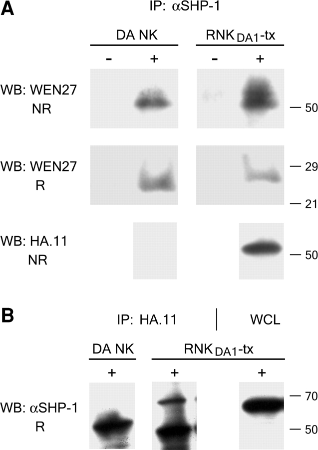
Figure 6.
KLRE1 is associated with SHP-1. Freshly isolated NK cells from DA rats (DA NK) and RNKDA1 cells transfected with a KLRE1-HA expression construct (RNKDA1-tx) were analyzed by immunoprecipitation with anti-SHP-1 (αSHP-1) (A) or anti-HA antibody (HA.11) (B). (A) Pervanadate stimulated (5 min at 37°C) (+) and unstimulated (−) cells were lysed in 1% digitonin buffer and immunoprecipitated with αSHP-1. The immune complexes were separated by SDS-PAGE under nonreducing (NR) and reducing (R) conditions respectively, blotted onto a membrane, and probed with WEN27 or anti-HA antiserum. Equal amounts of immunoprecipitated SHP-1 were verified by Western blotting using anti–SHP-1 antibodies (unpublished data). (B) Western blots of HA.11 immunoprecipitates from pervanadate stimulated cells probed with αSHP-1. The right lane with whole cell lysate (WCL) shows the position of SHP-1 (∼70 kD). The ∼50 kD band in the HA.11 immunoprecipitate lanes represents the Ig heavy chain of the anti-HA antibodies (SDS-PAGE performed under reducing conditions).

