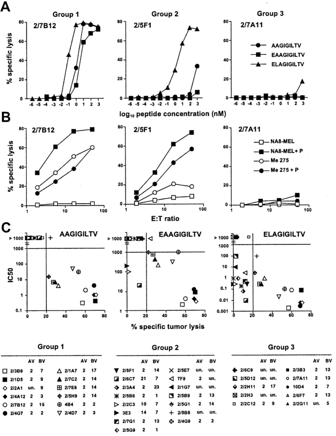
Figure 2.
Functional avidity of antigen recognition, fine specificity, and tumor-reactivity of ex vivo sorted A2/Melan-A multimer+ CD8+ T cell clones. Data are shown for one representative clone per group. (A) Clonal populations were tested for peptide recognition in chromium release assay using T2 cells as targets at a lymphocyte to target ratio of 10:1 in the presence of serial dilutions of the indicated peptide. (B) Tumor recognition was similarly assessed as the indicated lymphocyte to target cell ratios by using as target cells tumor cell lines Me275 (A2+, Melan-A+) and NA8-MEL (A2+, Melan-A−) in the absence or in the presence of peptide Melan-A 26–35 A27L (1 μM). (C) Correlation between avidity of antigen recognition and tumor reactivity of A2/Melan-A multimer+ clonal populations. Data obtained from the experiments illustrated in A and B are shown for 37 A2/Melan-A multimer+ clonal populations. The nM concentration of the indicated peptide which was required to obtain 50% of maximal lysis in peptide titration experiments (IC50) are shown in y-axis. The percent specific lysis on the Melan-A+ A2+ tumor line Me 275 obtained at an effector to target cell ratio of 50/1 in the absence of exogenously added peptide is shown in x-axis. Values >20% (bar on the X-axis) were considered as significant. Variable α and β (AV and BV) chain region usage of each clone is indicated. Un., unknown.

