
Figure 2.
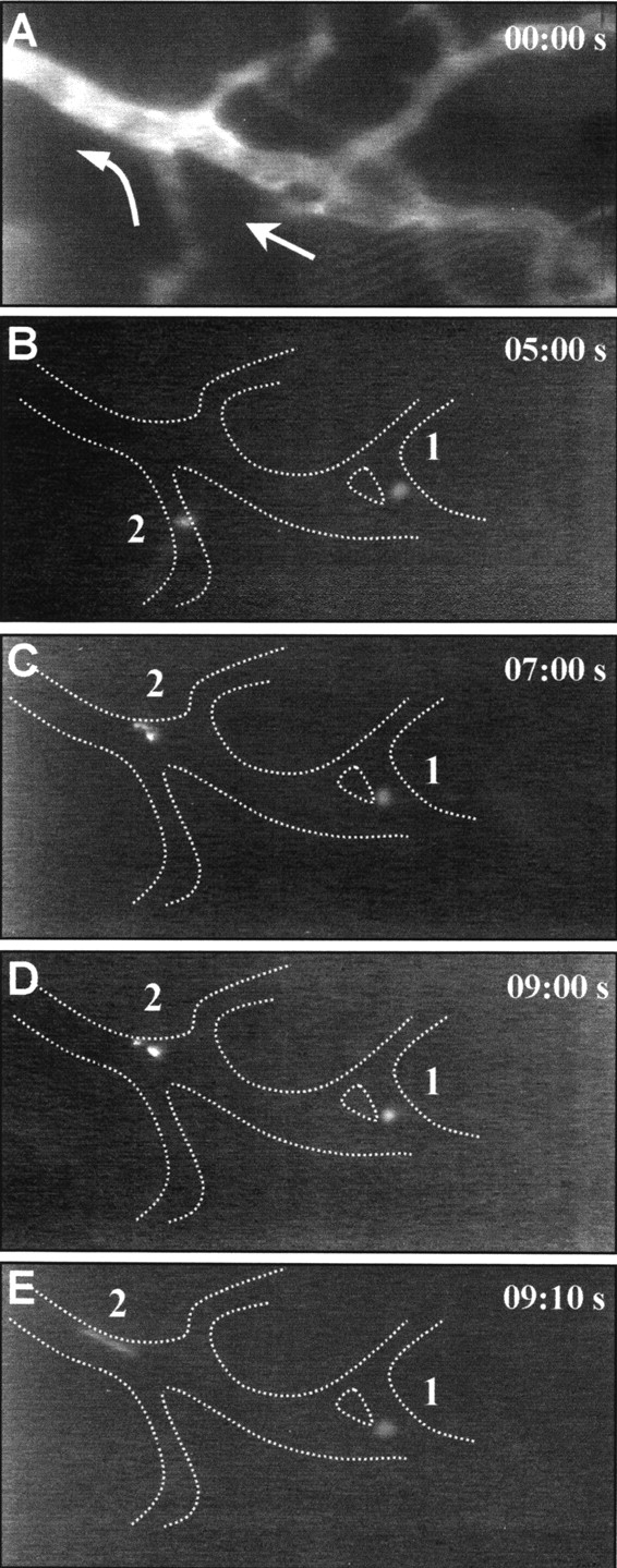
Circulating eEPCs interact with the tumor endo-thelium. (A) Tumor blood vessels before cell injection after contrast enhancement with FITC-conjugated dextran. Arrows indicate direction of microvascular blood flow. (B–E) Intravital microscopic sequence of two DiI-labeled eEPCs (1 and 2) interacting with the vessel wall of the identical vascular segment indicated in A. Cells adhere either permanently (1) or temporarily (2) to the endothelium. Numbers depict sequential time points in seconds (top right).