Figure 8.
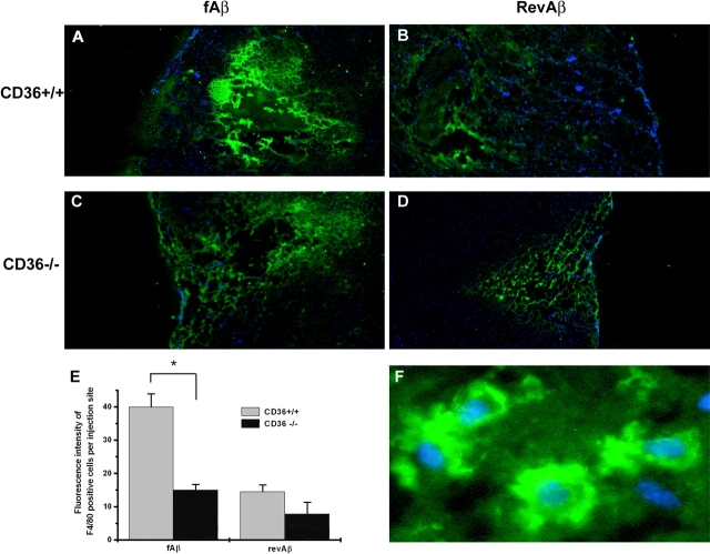
Intracerebral fAβ-induced recruitment of microglia is CD36 dependent. CD36+/+ (A and B) or CD36−/− (C and D) mice stereotaxically microinjected with 2 μg fAβ (A and C) or revAβ (B and D) into opposite hemispheres. 48 h later, mice were killed and 20-μm frozen sections were stained with F4/80 (green) and DAPI (blue; original magnification, ×4). The fluorescence intensity of F4/80 staining representing the number of microglia per injection site was quantified by digital imaging analysis (E). A higher magnification image showing details of cytoplasmic F4/80 and nuclear DAPI staining is shown in F. Each data point is the average ± SEM of five different sections per mouse and is a representative of five to seven mice per genotype with similar results. *, P < 0.005.

