
Figure 1.
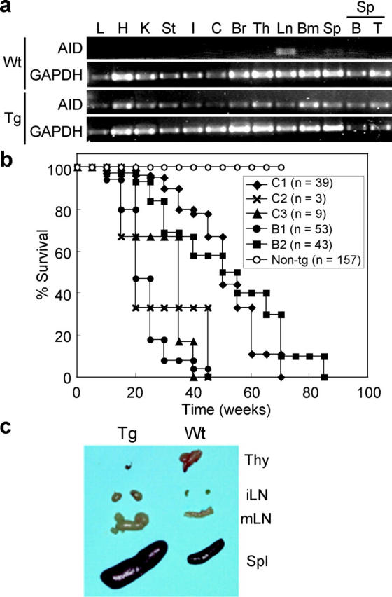
Early death and enlargements of lymphoid organs in AID Tg mice. (a) RT-PCR analysis of AID expression in wild-type and Tg tissues. Young (12 wk old) and healthy mice were used for analysis. GAPDH is an internal control. L, lung; H, heart; K, kidney; St, stomach; I, small intestine; C, colon; Br, brain; Th, thymus; Ln, mesenteric lymph nodes; Bm, bone marrow; Sp, spleen; B, purified splenic B cells; T, purified splenic T cells. No amplification was observed in RT(−) samples. (b) Survival curves of five independent lines of AID Tg mice and wild-type control. C1–3, Tg lines on C57BL/6 × (C57BL/6 × C3H/He) background; B1 and 2, Tg lines on C57BL/6 background. (c) Thymus (Thy), inguinal (iLN), and mesenteric (mLN) lymph nodes and spleen (Spl) from a 16-wk-old AID Tg mouse and its littermate control.