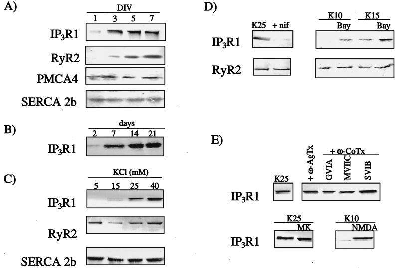
Figure 1.
Characterization of intracellular Ca2+ channels in developing granule cells by Western immunoblotting. In all cases, 25 μg of crude membrane proteins were separated and immunoblotted with isoform-specific antibodies. (A) Developmental profile in cerebellar granule cells of IP3R1, RyR2, PMCA4, and SERCA2b. DIV, days in vitro. (B) Developmental profile of IP3R1 in developing cerebellum. Days shown are days after birth. (C) Effect of different concentrations of KCl on the expression of IP3R1, RyR2, and SERCA2b. KCl concentrations were adjusted 1 day after plating, and cells were harvested on the fifth day. (D) Effect of agents acting on the L type Ca2+ channel on the expression of IP3R1. nif, nifedipine, 10 μM; BayK, BayerK8644, 1 μM. (E) Effect of agents acting on non-L type Ca2+ channels on IP3R1 expression. Agatoxins and ω-conotoxins all at 1 μM; MK, MK806, 10 μM; NMDA, 130 μM.

