
Figure 2.
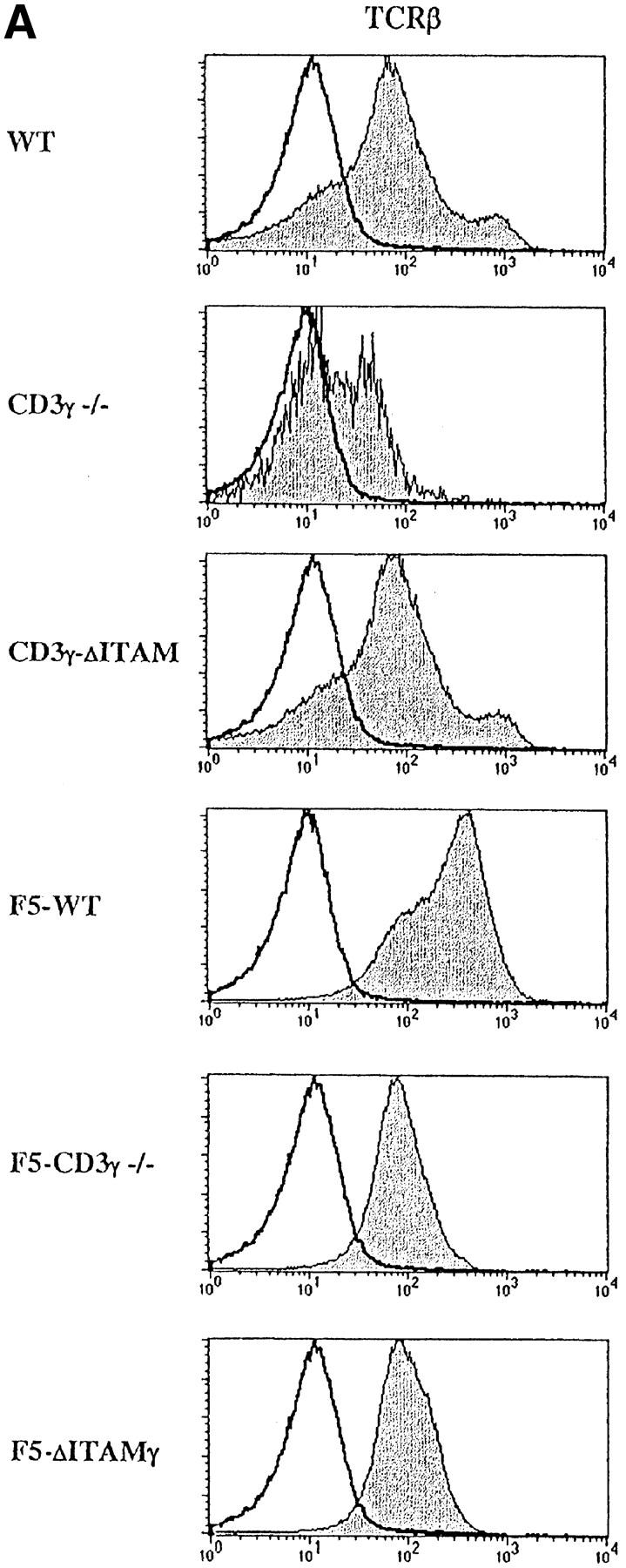
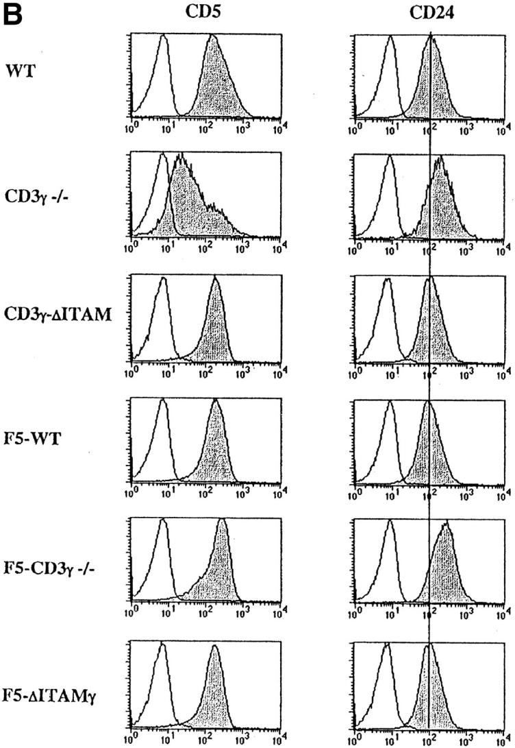
Analysis of TCR-, CD5-, and CD24- expression on DP thymocytes. Expression of cell surface TCRβ (A), CD5 and CD24 (B) (gray), or an irrelevant mAb (white) was analyzed within the electronically gated CD4+CD8+ DP compartment derived from 6–8-wk-old WT, CD3γ−/−, CD3γ–ΔITAM, F5–WT, F5–CD3γ−/−, and F5–ΔITAMγ mice.