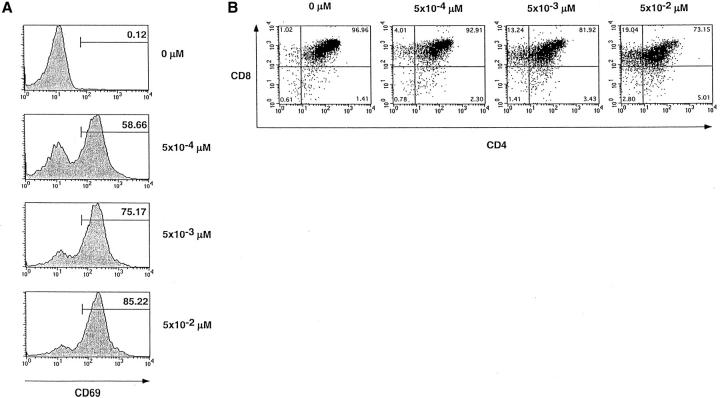
Figure 5.
CD69 upregulation and induction of CD8 SP thymocytes. Representation of the actual flow cytometry observations from which the percentage response data in Figs. 4 D and 6 are derived. Sorted F5–ΔITAMγ DP thymocytes were stimulated overnight in dispersed cultures with graded concentrations of the agonist peptide ASNENMDAM, and viable cells were stained for (A) CD69 or (B) CD4 versus CD8.

