
Figure 6.
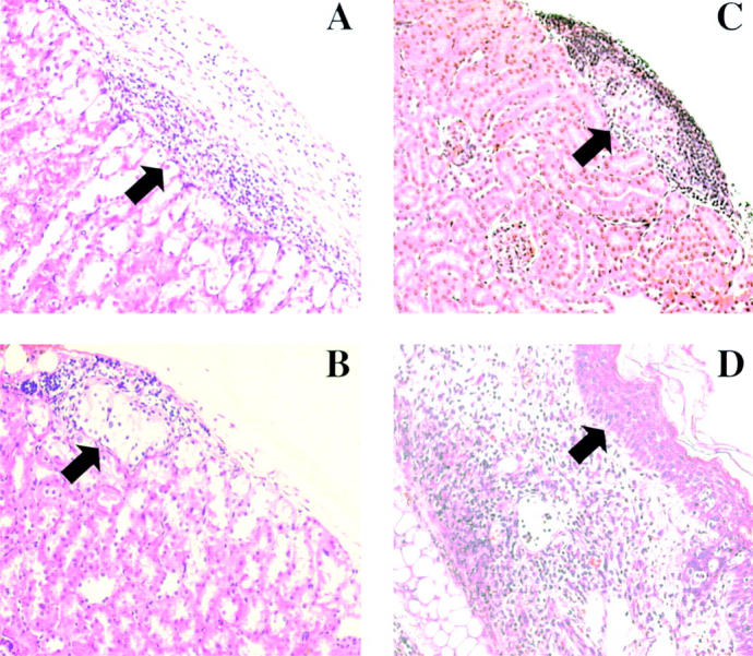
Graft histology. (A) Islet allograft transplanted into wild-type host harvested at the time of rejection (BG > 300 mg/dL). (B) Long surviving (>100 d) islet allograft removed from CD103−/− host as shown in Fig 1 C. (C) Islet allograft transplanted into CD103−/− host and harvested at the time of rejection in wild-type hosts (day 14 after transplant). (D) Skin allograft transplanted into wild-type host harvested at the time of rejection (day 8 after transplant). Arrows in A–C mark the position of islet allografts under the renal subcapsule; the arrow in D marks the postion of the graft epidermis. Data shown are H&E staining of paraffin-embedded sections.