
Figure 1.

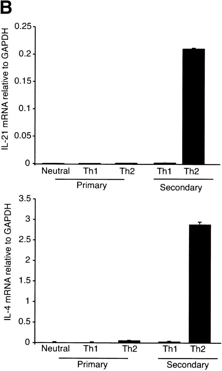


IL-21 is a Th2 cytokine. (A) Thp cells were cultured under Th1 and Th2 skewing conditions for 6 d. The cells were left resting (−) or restimulated with PMA/Ionomycin (P+I) for 4 h. Similar results were observed for 6 and 24 h after stimulation (unpublished data). RNA was purified and assessed for cytokine expression by Northern blot analysis. The results shown are representative of three independent experiments. (B) Thp cells were cultured under neutral, Th1, and Th2 skewing conditions. RNA was purified 24 h after primary and secondary anti-CD3 stimulation. Cytokine expression was assessed in duplicate by RealTime PCR and shown relative to GAPDH. (C) Thp cells were cultured under Th1 and Th2 skewing conditions for 5 d. IL-4 or IFN-γ were added to indicated cultures 24 h before secondary stimulation with anti-CD3. RNA was purified 24 h after secondary stimulation and IL-21 expression was assessed in duplicate and shown relative to GAPDH by RealTime PCR. (D) Cohorts of eight BALB/c and C57BL/6 mice were infected with L. major in hind footpads. After 6 wk CD4+ T cells from draining lymph nodes were purified and stimulated with anti-CD3. RNA was purified 6 h after stimulation and cytokine expression was assessed relative to GAPDH by RealTime PCR.