Figure 5.
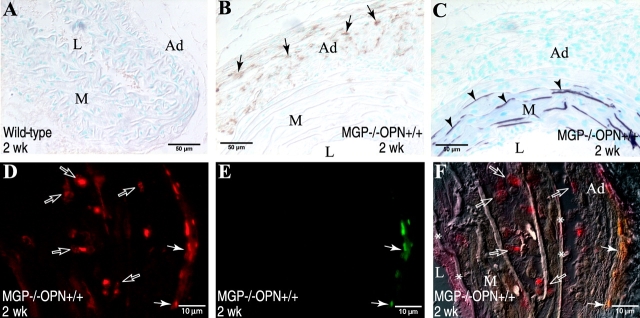
Macrophages are not the predominant cells to synthesize OPN in calcified aortas of MGP × OPN mutant mice. (A and B) Aortas of 2-wk-old MGP × OPN mutant mice were stained immunohistochemically for macrophages using streptavidin-conjugated peroxidase. Note the recruitment of macro-phages into (B) adventitia of calcified arteries (arrows), but not in (A) normal vessels. (C) Adjacent section stained for mineral (arrowheads) by Von Kossa. Colocalization of macrophages and OPN was further determined by double immunohistochemical staining using fluorophore-conjugated secondary antibodies. (D) Rhodamine-OPN, (E) Cyanine-BM8, and (F) Overlay of D and E onto a differential interference contrast photo of the same field. Note OPN+ cells (red in D and red and yellow in F) were present mostly in the medial layer where macrophages (green in E and green and yellow in F) were absent. Arrows, macrophages; open arrows, OPN-expressing cells except macrophages; arrowheads, mineral; L, lumen; M, media; Ad, adventitia; *, internal and external elastic laminae.

