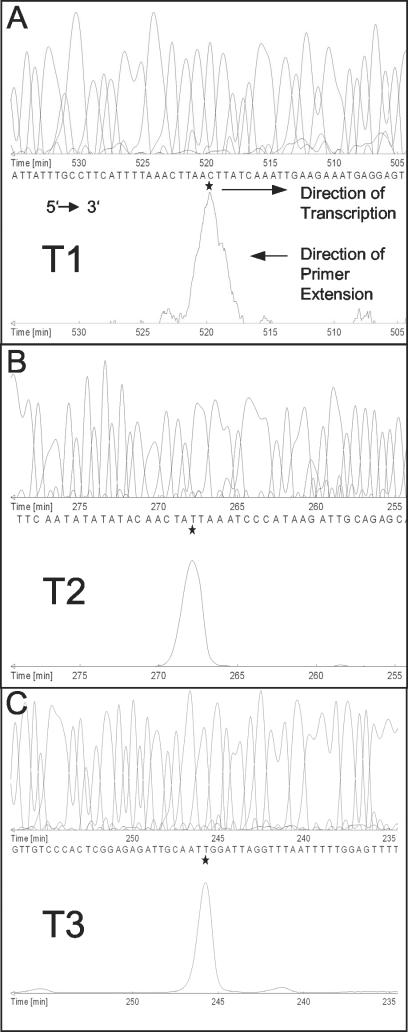
FIG. 2.
Mapping of the 5′ ends of three sae transcripts by fluorescence-based primer extension analysis. The precise base mapping was done by comparing the migration of the extended product with a parallel sequencing reaction primed by an identical Cy5-labeled oligonucleotide. The sequence and product traces are given in complement reverse order, thus corresponding to the 5′-to-3′ orientation. The initiation start nucleotide of the mRNA is marked by a star. The sae mRNAs of 3 (A), 2.4 (B), and 2 (C) kb are labeled T1, T2, and T3, respectively.

