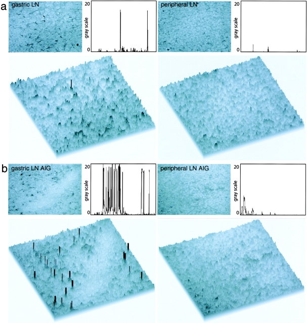
Figure 6.
Quantitative immunohistochemical analysis of H+/K+-ATPase in LN sections of untreated animals and animals with AIG. (a) Anti–H+/K+-ATPase FITC-stained LN sections of untreated BALB/c mice were incubated with secondary anti-FITC HRPO Ab and developed using enhanced diaminobenzidine substrate. Pictures were taken with the bright field setting of the microscope (top) and additionally analyzed using the public domain NIH Image program. Single peaks represent positive staining for H+/K+-ATPase in the threshold histogram plot profile (top) and in the surface plot view (bottom) and can be detected in gastric LN, but not peripheral LN, sections of untreated animals. (b) In animals with AIG, an increase in the staining frequency for H+/K+-ATPase was observed in gastric LN (gastric LN AIG), but not in peripheral LN (peripheral LN AIG), sections.

