
Figure 1.


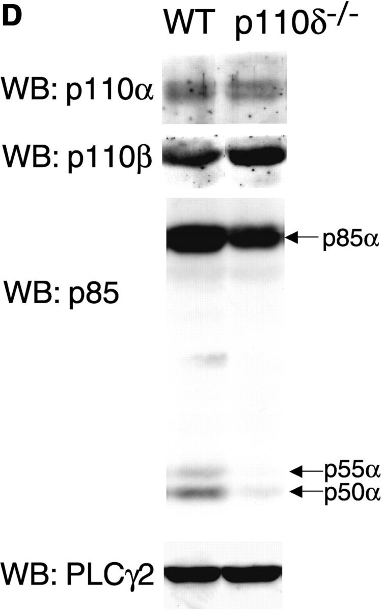


p110δ mutant B cells fail to produce PIP-3. (A) Gene targeting strategy. (B) Southern blot analysis of tail DNA from wild-type (+/+), heterozygous (+/−), and homozygous (−/−) mutant mice using probe A. (C) Western blot analysis of wild-type, heterozygous, and homozygous mutant thymocytes using antibody against p110δ. The blot was reprobed with anti–Vav-1 to demonstrate equal protein loading. (D) Western blot analysis of wild-type and homozygous mutant B-lymphocytes for levels of p110α, p110β, the p85/55/50α subunits of PI3-K, and PLCγ2. (E) BCR stimulated PIP-3 production in wild-type and mutant B cells. Wild-type is represented by black bars, mutant by white bars. (F) Time course of PIP-3 production in wild-type and mutant B cells stimulated with 10 μg/ml anti-IgM F(ab)2. In E and F, error bars represent the variance of triplicate determinations.